


Journal of Cardiology and Heart Failure
ISSN:
3139-6429 (Online)
DOI Prefix (Crossref): 10.67238
 Download PDF
ABSTRACT
Coronary computed tomography angiography (CCTA) has become an established noninvasive modality for excluding obstructive coronary artery disease; however, its diagnostic value extends beyond the coronary arteries. Careful evaluation of the entire acquired field of view may reveal clinically important extracardiac abnormalities that would otherwise remain undetected. We report the case of a 63-year-old woman who presented with persistent atypical chest pain and multiple cardiovascular risk factors, including hypertension, obesity, physical inactivity, and daily hookah smoking. Electrocardiography and transthoracic echocardiography were unremarkable. CCTA demonstrated a coronary artery calcium score of 0, CAD-RADS 0, and normal coronary arteries without evidence of atherosclerosis or stenosis, effectively excluding a cardiac cause of her symptoms. Unexpectedly, meticulous review of the extracardiac structures identified a 11.5 × 11.1 mm saccular aneurysm arising from the distal splenic artery. The patient had no abdominal symptoms or known predisposing conditions, including pancreatitis or portal hypertension, making the aneurysm a truly incidental finding. She was referred to vascular surgery, where conservative management with imaging surveillance was recommended because of the aneurysm's small size and low-risk features. This case emphasizes that clinically significant diagnoses may lie outside the primary indication for imaging. Systematic assessment of all structures included on CCTA can uncover potentially life-threatening vascular abnormalities, influence subsequent management, and prevent missed opportunities for early diagnosis. Greater awareness of incidental extracardiac findings is essential to maximize the diagnostic yield and clinical value of coronary CT examinations.
Keywords: Splenic Artery Aneurysm; Coronary Computed Tomography Angiography; Incidental Finding; Visceral Artery Aneurysm; Case Report
INTRODUCTION
Splenic artery aneurysm (SAA) is defined as a focal dilation of the splenic artery exceeding 50% of its normal diameter. It is the most common visceral artery aneurysm, accounting for approximately 60%–70% of reported visceral artery aneurysms. Although SAAs are uncommon, their widespread detection has increased with the use of cross-sectional abdominal and cardiovascular imaging, and they may be identified incidentally in asymptomatic patients [1-3].
A true SAA involves all three layers of the arterial wall, whereas a pseudoaneurysm results from disruption of the arterial wall with formation of a contained blood collection adjacent to the vessel. This distinction is clinically important because splenic artery pseudoaneurysms have a substantially higher risk of rupture and are generally managed differently from small, asymptomatic true aneurysms. Common risk factors for true SAA include female sex, advanced age, portal hypertension, pregnancy, and atherosclerotic disease. Pseudoaneurysms, in contrast, are most frequently associated with pancreatitis, abdominal trauma, or iatrogenic vascular injury [2-4].
Most SAAs are asymptomatic and are discovered incidentally during imaging performed for unrelated indications. When symptomatic, patients may present with abdominal or epigastric pain, nausea, vomiting, or gastrointestinal bleeding. Rupture is the most serious complication and may result in life-threatening intra-abdominal hemorrhage [2-4].
Management depends on aneurysm size, symptoms, morphology, interval growth, pregnancy or childbearing potential, and whether the lesion represents a true aneurysm or pseudoaneurysm. The 2020 Society for Vascular Surgery (SVS) clinical practice guidelines recommend treatment of nonruptured splenic artery true aneurysms >3 cm, those demonstrating interval enlargement, and symptomatic aneurysms. The guidelines also recommend treatment of splenic artery pseudoaneurysms regardless of size and of true aneurysms of any size in women of childbearing age. Endovascular treatment is commonly preferred when anatomically feasible because of its minimally invasive nature [5-6].
Coronary computed tomography angiography (CCTA) provides visualization not only of the coronary arteries and cardiac structures but also of adjacent thoracic and upper abdominal structures included within the acquired field of view. Clinically significant extracardiac findings are relatively common, and studies have demonstrated that restricting interpretation to the heart and coronary arteries may result in clinically relevant abnormalities being overlooked [7-9]. Therefore, systematic assessment of the complete CCTA field of view is important for identifying incidental abnormalities that may require further investigation or surveillance.
CASE PRESENTATION
A 63-year-old woman was referred to the cardiology outpatient clinic with a several-month history of intermittent chest pain associated with bilateral shoulder pain. The pain was atypical and non-exertional and was not associated with dyspnea, palpitations, syncope, or diaphoresis. She had not undergone previous cardiac evaluation.
Her medical history was significant for hypertension controlled with amlodipine and a pollen allergy. Her surgical history included cholecystectomy, appendectomy, and hysterectomy. She reported daily waterpipe (hookah) smoking and no regular physical exercise. There was no known history of diabetes mellitus, dyslipidemia, or established cardiovascular disease.
On physical examination, the patient was obese; otherwise, the cardiovascular, respiratory, and abdominal examinations were unremarkable. Her vital signs were within normal limits. A 12-lead electrocardiogram demonstrated normal sinus rhythm without ischemic abnormalities. Transthoracic echocardiography showed normal cardiac structure and function without regional wall motion abnormalities.
Given her cardiovascular risk factors and persistent chest discomfort, coronary computed tomography angiography (CCTA) was performed. The examination demonstrated a coronary artery calcium (CAC) score of 0 and a CAD-RADS score of 0, with no evidence of coronary atherosclerotic plaque or luminal stenosis. The coronary arteries demonstrated normal anatomy with right-dominant circulation. The cardiac chambers, cardiac valves, pericardium, and thoracic aorta were unremarkable.
During systematic assessment of the extracardiac structures included in the CCTA field of view, an incidental saccular aneurysm arising from the distal splenic artery was identified. The aneurysm measured approximately 11.5 × 11.1 mm (Figure 1). The lesion was located in the distal splenic artery and had imaging characteristics consistent with a true aneurysm rather than a pseudoaneurysm. There was no reported history of pancreatitis, abdominal trauma, or recent iatrogenic vascular injury to suggest a pseudoaneurysm. No additional significant extracardiac abnormalities were identified within the visualized field.
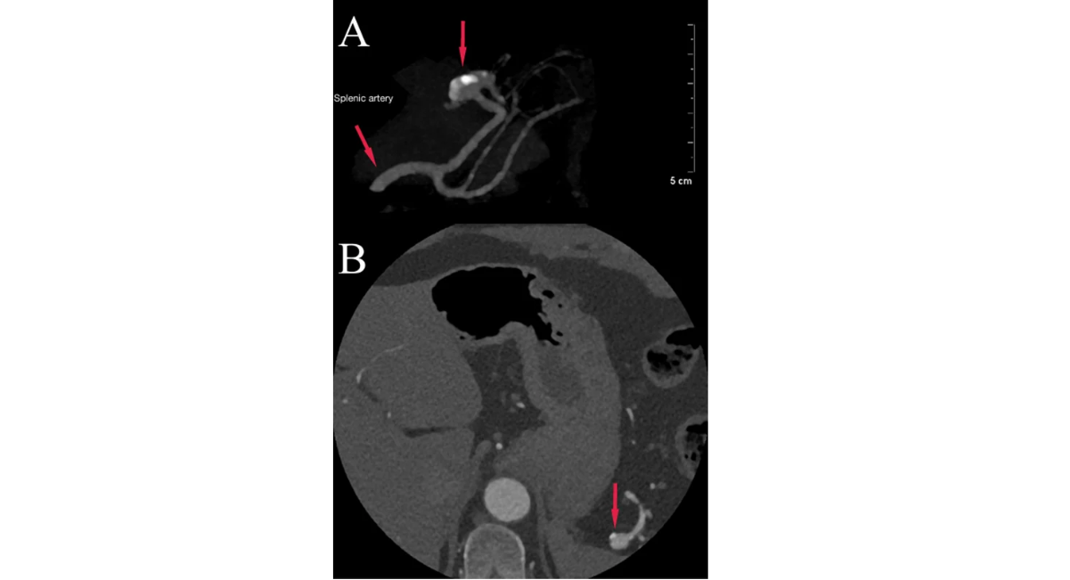
Figure 1: (A) Three-dimensional maximum intensity projection reconstruction demonstrating a saccular aneurysm (arrow) arising from a distal branch of the splenic artery. (B) Axial contrast enhanced CT image at the level of the upper abdomen from the same coronary CT angiography study, showing the aneurysm in cross section (arrow).
The patient was referred to the vascular surgery service for further evaluation of the incidentally detected splenic artery aneurysm. Given its small size, asymptomatic presentation, and absence of high-risk clinical features, conservative management with imaging surveillance was recommended. The patient was reassured that her cardiac evaluation demonstrated no evidence of coronary artery disease to explain her presenting chest pain.
DISCUSSION
We report the case of a 63-year-old woman with an incidentally detected distal splenic artery aneurysm measuring 11.5 × 11.1 mm, identified during coronary computed tomography angiography (CCTA) performed for the evaluation of atypical chest pain. Splenic artery aneurysm is the most common visceral artery aneurysm and is frequently detected incidentally with the increasing use of cross-sectional imaging [2-4].
The present case illustrates two clinically relevant aspects of CCTA: its ability to exclude coronary artery disease in patients with atypical chest pain and its potential to identify clinically important extracardiac abnormalities. In this patient, the CAC score was 0 and the CAD-RADS score was 0, with no evidence of coronary atherosclerosis or obstructive coronary artery disease. Careful review of the complete acquired dataset nevertheless revealed a vascular abnormality outside the primary cardiac field of interest.
The distinction between a true SAA and a pseudoaneurysm is particularly important because their etiologies, natural histories, and management differ. True aneurysms involve the intact arterial wall and are associated with factors such as portal hypertension, pregnancy, female sex, and advanced age. Splenic artery pseudoaneurysms are more commonly associated with pancreatitis, trauma, and iatrogenic injury and carry a higher risk of rupture. [2-4]. In the present case, the lesion was characterized as a true aneurysm based on its imaging appearance and the absence of a clinical history suggestive of pseudoaneurysm.
The aneurysm was located in the distal splenic artery rather than a separate distal branch. This anatomical description is used consistently throughout the manuscript because precise localization is relevant to both radiological characterization and potential future endovascular planning.
The 2020 SVS clinical practice guidelines recommend intervention for nonruptured splenic artery true aneurysms >3 cm, aneurysms demonstrating interval enlargement, and symptomatic aneurysms. Treatment is also recommended for splenic artery pseudoaneurysms regardless of size and for true aneurysms of any size in women of childbearing age [5]. The present lesion measured approximately 1.15 cm in its largest dimension, was asymptomatic, and was not associated with documented high-risk features. Therefore, its size was substantially below the SVS threshold for elective treatment of a nonruptured true splenic artery aneurysm.
Accordingly, the lesion was considered to have a relatively low-risk profile in the context of this patient. This designation does not imply that the aneurysm carries no risk; rather, it reflects its small size, asymptomatic presentation, apparent true-aneurysm morphology, and absence of recognized indications for immediate intervention. Conservative management with vascular specialist assessment and interval imaging surveillance was therefore appropriate. Surveillance allows assessment for interval enlargement or development of new clinical or morphological features that could alter the management strategy [5-6].
The clinical significance of this incidental finding extends beyond the aneurysm itself. Although the patient underwent CCTA because of chest pain, the coronary findings did not provide an explanation for her symptoms. The same examination identified a vascular abnormality with potential clinical consequences that would otherwise have remained undetected. Systematic review of extracardiac structures is therefore an important component of CCTA interpretation. A systematic review of 13 studies including 11,703 patients reported extracardiac findings in approximately 41% of CCTA examinations, with clinically significant findings in approximately 16% [7]. In another large study of 6,920 patients, 16.2% had extracardiac findings requiring therapy, additional workup, or follow-up, and a limited review strategy missed a substantial proportion of clinically relevant findings [8].
These observations emphasize the educational value of the present case. CCTA should not be interpreted solely as a coronary artery examination. The acquired field of view may include portions of the lungs, mediastinum, thoracic vessels, upper abdomen, and other structures, and careful assessment of these areas can reveal clinically relevant abnormalities [7-9]. In the current case, systematic evaluation of the available upper abdominal structures enabled recognition of an asymptomatic splenic artery aneurysm, prompting appropriate specialist referral and surveillance.
CONCLUSION
This case highlights the importance of systematically assessing the complete field of view obtained during coronary computed tomography angiography. Although CCTA was performed to evaluate atypical chest pain and demonstrated no evidence of coronary artery disease, careful review of the extracardiac structures revealed an incidental small distal splenic artery aneurysm. Recognition of such findings can facilitate appropriate specialist referral, risk assessment, and imaging surveillance. Clinicians interpreting CCTA should therefore remain attentive to extracardiac structures within the acquired field of view, as incidental vascular abnormalities may have important implications for subsequent patient management.
REFERENCES
- Kassem MM, Gonzalez L. Splenic artery aneurysm. In: StatPearls [Internet]. Treasure Island (FL): StatPearls Publishing; 2023 Jul 17. [Google Scholar] [PubMed]
- Al-Habbal Y, Christophi C, Muralidharan V. Aneurysms of the splenic artery—a review. Surgeon. 2010;8(4):223-231. [Crossref] [Google Scholar] [PubMed]
- Berceli SA. Hepatic and splenic artery aneurysms. Semin Vasc Surg. 2005;18(4):196-201. [Crossref] [Google Scholar] [PubMed]
- Pasha SF, Gloviczki P, Stanson AW, Kamath PS. Splanchnic artery aneurysms. Mayo Clin Proc. 2007;82(4):472-479. [Crossref] [Google Scholar] [PubMed]
- Chaer RA, Abularrage CJ, Coleman DM, Eslami MH, Kashyap VS, Rockman C, et al. The Society for Vascular Surgery clinical practice guidelines on the management of visceral aneurysms. J Vasc Surg. 2020;72(1S):3S-39S. [Crossref] [Google Scholar] [PubMed]
- Lim HJ. A review of management options for splenic artery aneurysms and pseudoaneurysms. Ann Med Surg (Lond). 2020;59:48-52. [Crossref] [Google Scholar] [PubMed]
- Karius P, Schuetz GM, Schlattmann P, Dewey M. Extracardiac findings on coronary CT angiography: a systematic review. J Cardiovasc Comput Tomogr. 2014;8(3):174-182.e1-e6. [Crossref] [Google Scholar] [PubMed]
- Johnson KM, Dennis JM, Dowe DA. Extracardiac findings on coronary CT angiograms: limited versus complete image review. AJR Am J Roentgenol. 2010;195(1):143-148. [Crossref] [Google Scholar] [PubMed]
- Aglan I, Jodocy D, Hiehs S, Soegner P, Frank R, Haberfellner B, et al. Clinical relevance and scope of accidental extracoronary findings in coronary computed tomography angiography: a cardiac versus thoracic FOV study. Eur J Radiol. 2010;74(1):166-174. [Crossref] [Google Scholar] [PubMed]

Article Processing Timeline
| Stage 1 |
Initial Quality & Plagiarism Check The manuscript is reviewed for journal scope, basic submission requirements, ethical compliance, and similarity/plagiarism concerns. |
| Stage 2 |
Editorial Assessment & Peer Review Manuscripts that pass the initial screening undergo editorial assessment and independent peer review in accordance with the journal's peer-review policy. |
| Stage 3 |
Revision & Editorial Decision Authors may be requested to revise their manuscript based on reviewer and editor comments. A final editorial decision is made after the required review and revision stages are completed. |
| Stage 4 |
Production & Publication Accepted manuscripts proceed to production, author proofing, final quality checks, and online publication. |
EDITORIAL & PUBLICATION INFORMATION
| English | Publication Language |
| Single-Blind | Peer-Review Model |
| 17% | Acceptance Rate |
| <18% | Similarity Screening Guideline |
| Open Access | Access Model |
All manuscripts undergo editorial assessment and originality screening as part of the journal's evaluation process. The acceptance rate shown is based on journal-level editorial data and may change over time. Similarity reports are assessed editorially and are not interpreted solely on the basis of a numerical similarity score.
Why Publish with us?
Ethics & Policies
Editorial & Management
Useful Links
Journal Highlights
Open Access Journals
Journal Flyer