Journal of Nanoscience and Nanomaterials
ABSTRACT
Sonoluminescence (SL) is a phenomenon in which transient flashes of light are emitted from gas-filled microbubbles when subjected to high-intensity ultrasonic irradiation. The conversion of acoustic energy into optical emission occurs during rapid cavitation and bubble collapse, generating localized regions of extreme temperature and pressure. Over the past decades, this phenomenon has transitioned from a physical curiosity to a scientifically relevant tool with applications in Chemistry, biomedical imaging, therapy, nanotechnology, and chemical engineering. In biomedical context, SL has gained importance due to its ability to generate light within tissues without the need for external optical penetration. Ultrasound frequencies commonly employed for SL in biological media range from approximately 20 kHz to several mega Hertz, depending on the intended application and depth of tissue penetration. Low-frequency ultrasound promotes cavitation and stronger bubble collapse, while higher frequencies provide controlled oscillation and improved spatial targeting. The unique fusion between ultrasound, microbubble contrast agents, and nanomaterials has enabled the development of hybrid imaging platforms capable of producing localized luminescent signals in vivo. These signals facilitate deep tissue imaging, targeted drug activation, and monitoring of therapeutic responses. Additionally, sonoluminescence plays a role in sonodynamic therapy, antimicrobial strategies, and molecular imaging. The integration of nanotechnology, particularly nanobubbles and functionalized nanoparticles, has significantly enhanced SL intensity, signal specificity, and biomedical activity. Beyond biomedical applications, SL has been widely investigated in sonochemistry, environmental remediation, and nanomaterial synthesis. The extreme microenvironments generated during cavitation drive chemical transformations and structural modifications at the nanoscale. New insights are provided on the mechanisms of SL, its biomedical imaging capabilities, therapeutic applications, integration with nanotechnology, and broader scientific applications, highlighting ongoing global research and technological innovation in the service of humanity and advancement of knowledge.
Keywords: Sonoluminescence; Acoustic Cavitation; Ultrasonic Irradiation; Biomedical Imaging; Sonodynamic Therapy; Microbubble Contrast Agents; Nanotechnology in Medicine; Reactive Oxygen Species; Deep Tissue Imaging; Sonochemistry
INTRODUCTION
Sonoluminescence (SL) refers to the emission of transient light from microscopic gas bubbles in a liquid medium subjected to intense ultrasonic irradiation [1]. The phenomenon occurs when acoustic energy induces rapid oscillation and subsequent collapse of microbubbles, resulting in localized high temperatures and pressures that produce photon emission. These light flashes are typically extremely brief, occurring on nanosecond timescales, yet their represent a remarkable conversion of sound energy into optical radiation. Initially observed in laboratory settings as a physical curiosity, SL has evolved into an important field of study integrating physics, chemistry, materials science, and biomedical engineering. The ability of ultrasound to penetrate biological tissues safely and non-invasively has led researchers to explore SL as a mechanism for imaging, drug activation, and therapeutic applications including detection of cancer tumors in the early stage [2].
In biomedical science, ultrasound is already widely used for diagnostic imaging as it is widely exploited in chemistry for the synthesis of nanomaterials and for the acceleration of kinetics of reactions like transesterification and fermentation [9]. The coupling of ultrasonics with luminescent bubble dynamics provides a platform for enhancing imaging contrast, tracking biological processes, and triggering therapeutic effects. This convergence has stimulated global race into the exploitation of the biomedical potential of SL [10]. Representative image of the phenomena of SL depicting the emission of light by the implosive collapse of the gas filled microbubble in the liquid medium along the path of ultrasonic waves is shown in Fig. 1.
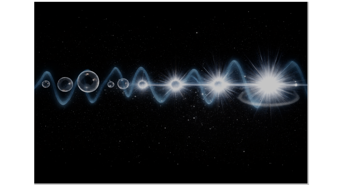
Figure 1: The optical emission of light in the nanosecond time scales from the implosive collapse of the gas filled micro bubble along the path of progress of the ultrasonic waves – The phenomena of microbubble sonoluminescence (MBSL).
Fundamental Mechanisms of Sonoluminescence
SL arises from acoustic cavitation, a process in which ultrasound generates oscillating bubbles within a liquid medium. These bubbles undergo cycles of expansion and contraction due to pressure variations in the acoustic field. At sufficiently high acoustic intensities, the bubbles collapse violently, producing localized extreme conditions characterized by high temperature, pressure, and rapid energy release. During collapse, the compression of gas within the bubble leads to excitation of atoms and molecules. As these excited states relax, photons are emitted, resulting in a flash of light. The emitted light spectrum can vary depending on gas composition, liquid medium, and acoustic parameters. The phenomenon can occur as single-bubble sonoluminescence or multi-bubble sonoluminescence, each with distinct dynamics and emission characteristics [11]. Key factors influencing SL include acoustic frequency, pressure amplitude, bubble (cavity) size, gas composition, and surrounding fluid properties. These parameters determine the efficiency of energy conversion and the intensity of emitted light. Understanding these mechanisms is critical for biomedical applications, where precise control over acoustic and optical effects is necessary.
SL originates from the process of acoustic cavitation, in which ultrasonic waves propagate through a liquid medium and induce the formation, oscillation, and eventual collapse of gas-filled microbubbles. The alternating high-pressure and low-pressure cycles generated by the acoustic field cause these microbubbles to undergo rapid expansion and compression. When the acoustic intensity surpasses a critical threshold, the bubbles become dynamically unstable and collapse violently. This implosion leads to the concentration of energy within an extremely small volume, producing localized conditions of elevated temperature and pressure. The rapid compression of the gaseous contents within the bubble is considered the primary driving factor responsible for the generation of light emission during the cavitation event. At the moment of collapse, the internal environment of the microbubble undergoes extreme thermodynamic changes, including adiabatic heating, ionization of gas molecules, and formation of transient plasma-like states. These conditions result in excitation of atoms and molecules, followed by radiative relaxation that manifests as the emission of photons. The spectral characteristics and intensity of the emitted light are influenced by several factors, including the composition of the gas within the bubble, the properties of the surrounding liquid medium, and the acoustic parameters applied. The phenomenon can occur as single-bubble sonoluminescence, where a stable oscillating bubble produces periodic light emission, or as multi-bubble sonoluminescence, where collective cavitation events generate diffuse luminescent output. The efficiency and dynamics of SL are governed by the interplay between acoustic pressure amplitude, frequency, bubble radius, and fluid viscosity. Lower ultrasonic frequencies generally promote stronger cavitation and more energetic collapse, while higher frequencies allow controlled oscillatory behaviour and spatial precision. Surface tension and dissolved gas concentration further influence bubble stability and emission characteristics. Contemporary mechanistic models suggest that energy focusing during implosion, shockwave formation, and rapid electron transitions collectively contribute to photon generation. Thus, SL represents a complex coupling of fluid dynamics, thermodynamics, and photo-physical processes that together enable the conversion of acoustic energy into optical radiation within microscopic environments within the time scales ranging from micro to pico seconds. The biomedical performance of SL arises from the interplay of acoustic, thermal, and photonic processes. Bubble collapse produces localized hotspots that generate light and reactive species, influencing biological molecules and cellular structures. These effects enable imaging, therapy, and molecular activation. Interactions with biological tissues depend on acoustic parameters and material properties. Controlled cavitation ensures targeted effects while minimizing tissue damage. Advances in acoustic engineering and nanotechnology are improving precision and safety.
Applications of Sonoluminescence
Sonoluminescence in Biomedical Imaging
SL has emerged as a promising mechanism for enhancing biomedical imaging due to its ability to generate localized optical emissions deep within biological tissues. Unlike conventional optical imaging, which is limited by light scattering and absorption, SL enables internal light generation through ultrasound-driven cavitation. Ultrasound frequencies used for biomedical SL typically range between 0.1 MHz and 5 MHz. Lower frequencies in the kilohertz range are particularly effective for inducing cavitation and generating intense luminescent emissions, whereas higher mega Hertz frequencies are preferred for diagnostic imaging and controlled bubble oscillation. Acoustic pressure amplitudes and pulse durations also influence luminescence intensity and spatial resolution [12]. Microbubble contrast agents play a critical role in SL-based imaging. These gas-filled bubbles, commonly used in ultrasound diagnostics, act as cavitation nuclei. When exposed to acoustic waves, their oscillate and collapse, producing luminescent signals that can be detected and correlated with tissue structures. These signals enhance imaging contrast, enabling visualization of vascular networks, tumor microenvironments, and inflammatory regions. SL has also been explored for molecular imaging. Functionalized microbubbles and nanobubbles can be engineered to bind specific cellular receptors or biomarkers. Upon acoustic activation, localized luminescence indicates the presence of targeted molecules, facilitating disease detection at early stages. This approach has potential applications in oncology, cardiovascular imaging, and infection monitoring [13]. The integration of SL with photoacoustic imaging techniques further improves imaging performance. Combined acoustic and optical signals provide complementary information regarding tissue structure, oxygenation, and metabolic activity. Such hybrid imaging systems are being investigated globally for high-resolution, non-invasive diagnostics [14].
Therapeutic and Diagnostic Applications
SL plays a significant role in therapeutic interventions where ultrasound-induced cavitation and luminescent emission activate biochemical processes. One of the most prominent applications is sonodynamic therapy, which involves ultrasound-mediated activation of sensitizing agents to generate reactive oxygen species. These reactive species induce apoptosis and necrosis in diseased cells, particularly cancerous tissues. Typical therapeutic ultrasound frequencies range from 0.02 MHz to 1 MHz for cavitation-driven therapy, as lower frequencies facilitate bubble collapse and reactive species production. Higher frequencies in the mega Hertz range are employed for targeted and controlled therapeutic effects. SLcontributes by providing localized photon emission that enhances activation of sensitizing molecules within tissues. Drug delivery systems benefit from SL through ultrasound-triggered permeability changes in cell membranes. Acoustic cavitation temporarily disrupts biological barriers, allowing targeted delivery of drugs and genes. Luminescent signals generated during therapy provide real-time monitoring of drug release and therapeutic efficacy [15]. Antimicrobial and antiviral applications arise from the generation of reactive oxygen species and localized heating during cavitation. These effects disrupt microbial membranes, denature proteins, and inhibit viral replication. SL-mediated processes are being explored for sterilization of medical devices, treatment of biofilms, and infection control in clinical environments [16]. Diagnostic applications include monitoring tissue response to therapy and assessing vascular perfusion. Luminescent signals correlate with cavitation activity and provide feedback regarding treatment intensity and localization. This enables optimization of therapeutic protocols and improves patient safety. The mechanism and function of sonodynamic therapy operating on SL with potential to kill the multidrug resistant bacteria is shown in Fig. 2.
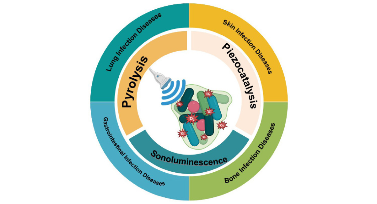
Figure 2: A schematic diagram of various mechanisms and applications of antibacterial sonodynamic therapy [Adapted with permission from 16].
Nano-sonoluminescence for multifunctional bioimaging, targeted drug delivery, and sonodynamic therapy
The integration of SL with nanotechnology has transformed its biomedical potential. Nanobubbles, nanoparticles, and functionalized contrast agents enhance cavitation efficiency and luminescent output. These materials act as acoustic sensitizers, improving control over bubble dynamics and energy transfer. Nanoparticles composed of silica, polymers, lipids, and carbon-based materials can be engineered to respond to ultrasonic stimulation. Their interaction with acoustic waves amplifies luminescent emissions and enables targeted imaging. Functionalized nanostructures carrying ligands or antibodies allow selective binding to diseased tissues, increasing diagnostic accuracy. Nanobubbles are generated at ultrasound frequencies typically between 0.5 MHz and 3 MHz, providing stable oscillation and controlled cavitation. Their small size allows penetration into microvascular and interstitial spaces, enabling imaging of deep tissues and tumor environments [17]. Hybrid nanoplatforms combining SL with photoluminescent or magnetic materials have been developed for theranostic applications. These systems provide simultaneous imaging, drug delivery, and therapy. Nanoparticles can also serve as carriers for therapeutic agents, releasing them upon acoustic activation. Energy transfer between acoustic waves and nanomaterials enhances generation of reactive oxygen species, enabling improved sonodynamic therapy. The integration of nanotechnology also improves signal sensitivity, reduces acoustic thresholds, and enhances biocompatibility. The persistence of the SL signal even after 10 minutes by integrating the ultrasound generated microbubbles with the nanocrystals of ZnO and lipase coated ZnO is shown in Fig. 3 [17]. A Schematic of the ZnO and ZnO-lipase nanocrystal’ structure and composition is shown in Fig. 3A. TEM image of ZnO nanocrystals is shown in Fig. 3 B. The TEM image of Lipase coated ZnO nanocrystals is shown in Fig. 3 C. A Schematic representation and the ultra high speed images of the ultrasound genic micron sized gas pockets entrapped at the ZnO surface’ coalesce into bigger micrometer bubbles stabilized by ZnO nanocrystals is shown in Fig. 3 D. Echo graphic contrast signal demonstrating the persistence of the SL signal in a sample tissue where in water (at time 0), ZnO suspension (at time 0 and at 5 min) and ZnO-lipase suspension (at 5 minutes and 10 minutes) were injected; echo graphic images (E) at time 0 min, (F) at time 5 min and (G) at time 10 min (Fig. 3 E, F, and G) [17].
Figure 3: Schematic illustration of sonoluminescence-assisted and ultrasound-based dual-mode imaging using hybrid nanocrystals (ZnO and Lipase coated ZnO)
Sonoluminescence for materials synthesis and environmental remediation
SL has significant applications outside the biomedical domain, particularly in chemistry, environmental science, and materials engineering. The extreme conditions generated during cavitation, including high temperatures and pressures, resulting in the SL drive chemical reactions and physical transformations. In sonochemistry, SL is associated with the formation of reactive radicals that initiate photochemical reactions. These processes are utilized in pollutant degradation, organic synthesis, synthesis of exotic nanomaterials and catalysis. Acoustic frequencies between 20 kHz and 100 kHz are commonly employed for chemical applications, as their produce strong cavitation effects [18]. Environmental remediation applications include water purification and degradation of persistent organic pollutants. SL enhances oxidation reactions that break down contaminants into less harmful compounds. This approach has been explored for wastewater treatment and industrial effluent management which is more significant in these days of rapid globalization that has resulted in population explosion, urbanization and severe deterioration of air, water and soil quality. In material’s science, SL contributes to synthesis of nanoparticles and nanostructured materials. The high-energy environment during bubble collapse promotes nucleation and growth of nanomaterials with unique properties. Acoustic parameters influence particle size, morphology, and crystallinity. Industrial applications include surface cleaning, emulsification, coatings and process monitoring. SL signals can be used as indicators of cavitation intensity and system efficiency. These processes are applied in microfabrication, pharmaceutical production, and chemical engineering [19]. Emerging research is exploring energy-related applications, including hydrogen production and catalytic reactions driven by cavitation. The ability of SL to generate localized extreme conditions provides opportunities for innovative technological developments across multiple scientific disciplines.
CHALLENGES AND FUTURE DIRECTIONS
Despite promising advances, challenges remain in translating sonoluminescence (SL) into routine clinical applications. Precise control of cavitation dynamics, reproducibility of luminescent signals, and safety considerations require further investigation. Understanding long-term biological effects and optimizing acoustic parameters are essential. Future research is expected to focus on hybrid imaging systems combining ultrasound, optical techniques, and nanotechnology. Personalized medicine approaches may utilize SL for targeted diagnostics and therapy. Advances in computational modeling and experimental techniques will enhance mechanistic understanding. For a more in depth knowledge of SL that can lead to path braking findings the readers are advised to consult the following literature [20-30].
Nanoscale Mechanisms and Material Design Considerations
Some vital mechanisms leading to the enhancement of SL phenomena due to the synergistic interaction of nanoparticles (ZnO, SiO2, CNDs, graphene, Ag, Au, rare earth based quantum dots include the enhancement of the cavitation, localization/concentration of the acoustic energy, intensification of the implosive collapse of the gas pockets, and optical and plasmonic coupling [31-34]. Nanoscale surface properties of materials does affect the cavitation dynamics and energy transfer resulting in the enhancement of SL. The unique synergy between the surface properties of nanoparticles and the acoustic (ultra sound) waves significantly improve the SL and this is attributed to the modification of the acoustic field, cavitation dynamics and the light emission processes in the presence of the nanoparticles (Fig. 4).
Figure 4: Synergistic interaction of nanoscale surfaces and ultrasound leading to enhanced sonoluminescence. Adapted with permission from Beilstein-Institute Open Science [35].
Nanoparticles serve as the heterogeneous nucleation centers for the cativation (Fig. 5).
Figure 5: Possible nanoscale surfaces that respond to ultrasound and the mechanisms for enhanced sonoluminescence. Adapted with permission from Beilstein-Institute Open Science [35].
As a result, the threshold for the formation of gas pockets is lowered with increased bubble formation resulting in the stable, sustainable and intense cavitation dynamics. Acoustic cavitation events and hence the frequency of the collapse of bubbles and emission of SL are enhanced by the surface properties of the nanoparticles like the high specific surface area, surface functional groups, surface plasmon resonance, surface heterogeneity and defect sites apart from many other surface features which are manifested at the nanoscale. These peculiar surface properties of the nanoparticles enable them to trap the dissolved gases creating the gas cavities under sonochemical irradiation upon whose collapse the phenomena of sonolumenscence is observed with increased intensity. Some ways in which the nanoparticle surfaces interact with ultrasound include, acoustic scattering, localized pressure gradients and thermoelastic vibraitons. Such interactions at the nanoscale surface lead to local amplification of pressure oscillations and increase the events of bubble collapse near the nanoscale surfaces. In the specific case of metallic nanoparticles like Au and Ag the ultrasonic waves excites the nano-mechanical vibrations resulting in the enhancement of local energy density around the nanoparticles, thereby increasing the bubble collapse intensity leading to stronger SL. Nanoparticles with optical properties like the semiconductors, plasmonic materials, carbon nanodots, rare earth containing nanomaterials, are often used for enhancing the SL (Fig. 5). A condensed version of the role of nanoparticles in enhancing the phenomena of SL and the mechanism thereof is highlighted in Table 1.
|
S.No |
Nanoparticles |
Mode of enhancement of Sonoluminescence |
Reference |
|
1 |
ZnO nanocrystals; Amine functionalized ZnO nanoparticles |
Decrease in cavitation threshold |
[32] |
|
2 |
Amimopropyl functionalized ZnO nanocrystals |
Formation of bigger cluster of bubbles and the resulting absorption/shielding of the collapsing events; lowering of threshold for the appearance of light emission |
[31] |
|
3 |
TiO 2 fractured nanoshells (TFNs) |
Inertial cavitation |
[42] |
|
4 |
Lipase coated ZnO nanocrustals |
Inertial cavitation |
[17] |
Table 1: Mechanism of Enhancement of Sonoluminescence by Nanoparticles
As the application domain of the phenomena is rapidly expanding, there is need for further clarification on the origins of the phenomena which need intense research [36-42]. Greater clarity into the mechanisms of enhancement of the SL phenomena in the presence of functionalized nanocrystals is awaited.
CONCLUSION
Sonoluminescence represents a unique fusion of acoustics, optics, nanotechnology and biomedical science. Its ability to generate localized light and reactive species through acoustic stimulation offers significant potential in chemical synthesis, imaging, therapy, and diagnostic monitoring. The integration of nanotechnology and advanced acoustic systems has expanded its applicability across biomedical and industrial domains. Ongoing global research continues to explore its mechanisms and applications, positioning sonoluminescence as a promising tool in next-generation biomedical technologies. With further refinement in control, safety, and integration, sonoluminescence is poised to contribute substantially to advances in imaging, targeted therapy, and translational medicine including the early diagnosis and imaging of cancer tumors. Many facts about the origin of the sonoluminescence are not yet publicly disclosed necessitating intense study on this phenomena.
ACKNOWLEDGMENTS
Thanks are due to Professor Deepak Nallaswamy for the state-of-the-art facilities at SIMATS. Gratefulness is due to Professor Nisha Chandru for the research facilities and resources. AGR thanks his parents Mr Ravichandran and Mrs Rajalakshmi for they steadfast support. Indebtedness is due to Dr Anandamurugan, librarian (in-charge), and the staff of the central library IIT Madras for the knowledge resources. Gratefulness is due to Mrs Saradhambal V, Superintendent, Central library IIT Madras for the facilities.
AUTHOR’S CONTRIBUTION
AGR wrote the original short communication. INP edited the manuscript and provided new insight. AG conceived and introduced the topic and provided the guidance and insight.
CONFLICTS OF INTEREST
The authors declare no conflicts of interest.
REFERENCES
- Maksimenko VV, Lushnikov AA, Zagaynov VA, Agranovski IE. Plasmon Mechanism of Sonoluminescence in Water. Russian Journal of Physical Chemistry A. 2025;99(12):3150-8. [Crossref] [Google Scholar]
- McLeod J, Bau L, Walton S, Katrinka Mavrak J, Lane R, Callan J, et al. Exploring the role of sonoluminescence in sonodynamic therapy. The Journal of the Acoustical Society of America. 2025;157(4_Supplement):A302-3. [Crossref] [Google Scholar]
- Indra Neel P, Gedanken A, Schwarz R, Sendersky E. Mild sonication accelerates ethanol production by yeast fermentation. Energy & fuels. 2012;26(4):2352-6. [Crossref] [Google Scholar]
- Tzhayik O, Pulidindi IN, Gedanken A. Forming nanospherical cellulose containers. Industrial & Engineering Chemistry Research. 2014;53(36):13871-80. [Crossref] [Google Scholar]
- Korzen L, Pulidindi IN, Israel A, Abelson A, Gedanken A. Single step production of bioethanol from the seaweed Ulva rigida using sonication. RSC advances. 2015;5(21):16223-9. [Crossref] [Google Scholar]
- Klein M, Varvak A, Segal E, Markovsky B, Pulidindi IN, Perkas N, et al. Sonochemical synthesis of HSiW/graphene catalysts for enhanced biomass hydrolysis. Green Chemistry. 2015;17(4):2418-25. [Crossref] [Google Scholar]
- Pulidindi IN, Gedanken A. The catalytic production of biofuels (biodiesel and bioethanol) using sonochemical, microwave, and mechanical methods. In Nontraditional Activation Methods in Green and Sustainable Applications 2021(pp. 171-239). Elsevier. [Crossref] [Google Scholar]
- Pulidindi IN, Gedanken A. Can biofuels alleviate the energy and environmental crisis? [Google Scholar]
- Pulidindi IN, Gedanken A. Employing novel techniques (Microwave and Sonochemistry) in the synthesis of biodiesel and bioethanol. In Production of Biofuels and Chemicals with Ultrasound 2014 (pp. 159-185). Dordrecht: Springer Netherlands. [Crossref] [Google Scholar]
- Canaparo R, Foglietta F, Giuntini F, Francovich A, Serpe L. The bright side of sound: Perspectives on the biomedical application of sonoluminescence. Photochemical & Photobiological Sciences. 2020;19(9):1114-21. [Crossref] [Google Scholar] [PubMed]
- Borisenok VA, Sedov SY. On the mechanisms of sonoluminescence in polar and nonpolar liquids. Physics of Atomic Nuclei. 2020;83(11):1575-84. [Crossref] [Google Scholar]
- He Y, Xing D, Yao Y, Tang Y, Tan S, Ueda KI. Enhanced sonoluminescence of tissues. In Biomedical Photonics and Optoelectronic Imaging 2000 (Vol. 4224, pp. 236-240). SPIE. [Crossref] [Google Scholar]
- He Y, Xing D. Sonoluminescence optical confocal scanning tomography of tissue. In Biomedical Optoacoustics II 2001 (Vol. 4256, pp. 249-255). SPIE. [Crossref] [Google Scholar]
- Xing D, He Y, Tang Y, Tan S. Sonoluminescence imaging and its biomedical applications. In Biomedical Photonics and Optoelectronic Imaging 2000 (Vol. 4224, pp. 196-200). SPIE. [Crossref] [Google Scholar]
- Beguin E, Shrivastava S, Dezhkunov NV, McHale AP, Callan JF, Stride E. Direct evidence of multibubble sonoluminescence using therapeutic ultrasound and microbubbles. ACS applied materials & interfaces. 2019;11(22):19913-9. [Crossref] [Google Scholar] [PubMed]
- Yi S, Gao Y, Yu L, Chen Y. Antibacterial sonodynamic nanomedicine: mechanism, category, and applications. Biomaterials Translational. 2025;6(1):24. [Google Scholar] [PubMed]
- Vighetto V, Pascucci E, Percivalle NM, Troia A, Meiburger KM, Van Den Broek MR, et al. Functional nanocrystal as effective contrast agents for dual-mode imaging: Live-cell sonoluminescence and contrast-enhanced echography. Ultrasonics sonochemistry. 2025;113:107242. [Crossref] [Google Scholar] [PubMed]
- Aghelmaleki A, Afarideh H, Cairós C, Pflieger R, Mettin R. Effect of mechanical stirring on sonoluminescence and sonochemiluminescence. Ultrasonics Sonochemistry. 2024;111:107145. [Crossref] [Google Scholar] [PubMed]
- Kang KM, Kim HW, Shim IW, Kwak HY. Syntheses of Specialty Nanomaterials at the Multibubble Sonoluminescence Condition. In ASME International Mechanical Engineering Congress and Exposition 2008 (Vol. 48760, pp. 101-103). [Crossref] [Google Scholar]
- Xu H, Glumac NG, Suslick KS. Temperature inhomogeneity during multibubble sonoluminescence. Angewandte Chemie International Edition. 2010;49(6):1079-82. [Crossref] [Google Scholar] [PubMed]
- Unnikrishnan CS, Mukhopadhyay S. Sonoluminescence as Quantum Vaccum Radiation. arXiv preprint quant-ph/9606016. 1996. [Crossref] [Google Scholar]
- Suslick KS, Flint EB. Sonoluminescence from non-aqueous liquids. Nature. 1987;330(6148):553-5. [Crossref] [Google Scholar] [PubMed]
- Suslick KS, Doktycz SJ, Flint EB. On the origin of sonoluminescence and sonochemistry. Ultrasonics. 1990;28(5):280-90. [Crossref] [Google Scholar] [PubMed]
- West CD, Howlett R. Timing of sonoluminescence flash. Nature. 1967;215(5102):727. [Crossref] [Google Scholar]
- Flint EB, Suslick KS. Sonoluminescence from nonaqueous liquids: emission from small molecules. Journal of the American Chemical Society. 1989 Aug;111(18):6987-92. [Crossref] [Google Scholar]
- Leighton TG, Pickworth MJ, Tudor J, Dendy PP. A search for sonoluminescence in vivo in the human cheek. Ultrasonics. 1990;28(3):181-4. [Crossref] [Google Scholar] [PubMed]
- Moss WC, Clarke DB, White JW, Young DA. Sonoluminescence and the prospects for table-top micro-thermonuclear fusion. Physics Letters A. 1996;211(2):69-74. [Crossref] [Google Scholar]
- Rao KR. Nuclear fusion using a table-top sonoluminescence device. Current Science. 2002;82(11):1317. [Google Scholar]
- Arakeri VH. Sonoluminescence and bubble fusion. Current science. 2003:911-6. [Google Scholar]
- Lahey Jr RT, Taleyarkhan RP, Nigmatulin RI. Sonofusion technology revisited. Nuclear Engineering and Design. 2007;237(15-17):1571-85. [Crossref] [Google Scholar]
- Vighetto V, Troia A, Laurenti M, Carofiglio M, Marcucci N, Canavese G, et al. Insight into sonoluminescence augmented by ZnO-functionalized nanoparticles. ACS omega. 2022;7(8):6591-600. [Crossref] [Google Scholar] [PubMed]
- Maksymov IS. Gas bubble photonics: Manipulating sonoluminescence light with fluorescent and plasmonic nanoparticles. Applied Sciences. 2022;12(17):8790. [Crossref] [Google Scholar]
- Song D, Xu W, Luo M, Zhang M, Wen H, Cheng X, et al. Influence of carbon nano-dots in water on sonoluminescence. Nanoscale. 2021;13(33):14130-8. [Crossref] [Google Scholar] [PubMed]
- Yang N, Li J, Yu S, Xia G, Li D, Yuan L, et al. Application of nanomaterial-based sonodynamic therapy in tumor therapy. Pharmaceutics. 2024;16(5):603. [Crossref] [Google Scholar] [PubMed]
- Fateh ST, Moradi L, Kohan E, Hamblin MR, Dezfuli AS. Comprehensive review on ultrasound-responsive theranostic nanomaterials: mechanisms, structures and medical applications. Beilstein Journal of Nanotechnology. 2021;12(1):808-62. [Crossref] [Google Scholar] [PubMed]
- Roberts PH, Wu CC. Structure and stability of a spherical implosion. Physics Letters A. 1996;213(1-2):59-64. [Crossref] [Google Scholar]
- Borisenok VA. Sonoluminescence: Experiments and models. Acoustical Physics. 2015;61(3):308-32. [Crossref] [Google Scholar]
- Seifritz W. A new way of tapping nuclear fusion energy? Ein neuer Weg zur Nutzbarmachung der Kernfusion? Internationale Zeitschrift fuer Kernenergie. 1996;41. [Google Scholar]
- Taleyarkhan RP, West CD, Cho JS, Lahey Jr RT, Nigmatulin RI, Block RC. Evidence for nuclear emissions during acoustic cavitation. Science. 2002;295(5561):1868-73. [Crossref] [Google Scholar] [PubMed]
- Sadighi-Bonabi R, Gheshlaghi M. Laser induced sonofusion: A new road toward thermonuclear reactions. AIP Advances. 2016;6(3). [Crossref] [Google Scholar]
- Xu Y, Butt A. Confirmatory experiments for nuclear emissions during acoustic cavitation. Nuclear Engineering and Design. 2005;235(10-12):1317-24. [Crossref] [Google Scholar]
- Jonnalagadda US, Su X, Kwan JJ. Nanostructured TiO2 cavitation agents for dual-modal sonophotocatalysis with pulsed ultrasound. Ultrasonics sonochemistry. 2021;73:105530. [Crossref] [Google Scholar] [PubMed]
Article Processing Timeline
| 2-5 Days | Initial Quality & Plagiarism Check |
| 25-35 Days |
Peer Review Feedback |
| 45-60 Days | Total article processing time |
Journal Flyer